Microtomography
X-ray microtomography (XMT) is a non-destructive evaluation technique that allows the internal structure of an object to be imaged by reconstructing the spatial distribution of the local linear X-ray absorption coefficients of the materials/phases contained within. This provides a virtual 3D representation of the internal architecture of an object from which two-dimensional (2D) cross-sectional slices can be extracted along the three orthogonal planes of an object. The 3D object can be converted into a microstructurally faithful 3D mesh suitable for Finite Element Modelling that can describe the geometry of each constituent.
The University of Manchester has embarked upon a bold investment aimed at creating the most comprehensive and capable 3D X-ray imaging capability anywhere in the world. The Manchester X-ray Imaging Facility is founded around two major pillars: the Henry Moseley X-ray Imaging Facility focussing on laboratory based tomography and the MXIF facility at the Research Complex at Harwell, where both laboratory and synchrotron based imaging are conducted.
Together these represent an investment of over £11M (£7M capital & £4M in staff) and an activity encompassing over 15 academics and over 100 users. It provides a window into structures from as 1 m to 10's of microns in size, with resolutions from 100's microns all the way down 50nm with the highest resolution machines in the UK. The unique in situ sample environment experimental rigs allow the resolution of processes occurring over timescales from years to under a second, making the imaging 4D.
The Manchester X-ray Imaging Facility provides openly available access under both academic and commercial arrangements to 7 complementary laboratory CT systems for non-destructive 3D imaging on millimetre to nanometre lengthscales. This is complimented by the Manchester-Diamond beamline at the Diamond Light Source synchrotron. The MXIF complements and extends the existing capabilities within the Department of Materials for the non-destructive investigation of materials across a very wide range of disciplines.
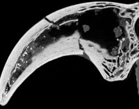
Given the spatial heterogeneity of most geological and palaeontological samples, and the highly variable scale of dynamic geological processes, the ability of x-ray CT to acquire non-destructive 3D and 4D imaging of a sample can be of great use in a wide range of geological and environmental fields. We have extensive experience on both laboratory scanning equipment and a many of the synchrotron imaging beam lines suitable for such samples.
Lead researchers
- Dr. Russell Garwood
- Prof. Phil Manning
- Prof. Philip Withers
- Dr. Tristan Lowe
- Prof. William Sellers
- Dr. Victoria Egerton
- Dr. Lee Margetts
- Dr. Charlotte Brassey
- Dr Thomas O'Mahoney
- Dr Mike Buckley